

Biomedical Imaging
Description
The multimodal platform perform advanced Imaging studies and sequential analyses of the same sample by means of both Magnetic Resonance Imaging (MRI) and µCT (Computed Microtomography). Notably, this approach offers the unique advantage to directly compare the images obtained by the two techniques, using the same software.
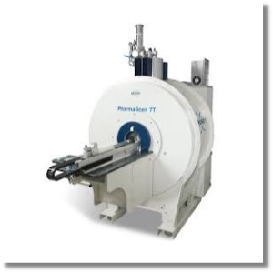
MRI allows to acquire a wide range of morphologic, metabolic and functional information from the nervous system and several other organs and tissues. In particular, the application of this technique is crucial for non invasive studies of tumors, edema, lesions, of the hearth (Cardio MRI), and for studies regarding the connectivity and the functions of the different areas activated within the nervous system (e.g. by means of Diffusion Tensor Imaging and Functional Imaging, fMRI). Moreover, using the MRI tomograph, it is possible to conduct Magnetic Resonance Spectroscopy (MRS) analyses, which retrieves the simultaneous and accurate quantification of several metabolites contained in tissues, such as N-acetylaspartate (NAA), Choline (Cho), Creatine (Cr), Myo-inosytol (mIns), Glutamate (Glu), Glutamine (Gln), Lactate (Lac), whose concetration can be severely altered in presence of pathologies , e.g. neurodegenerative diseases and tumors.
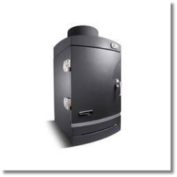
X-ray Computed Microtomography (microCT) provides the 3-dimensional internal structure of samples with a minimum pixel size of 2.8 μm. The 3D structure is the result of a complex reconstruction, obtained from a set of 2-dimensional shadow images of samples, produced by the X rays passing through them. MicroCT is optimal for the study of bones, to obtain highly detailed 3D reconstructions without compromising the subsequent genetic and histological analyses. The application of this technique, with the simultaneous usage of contrast agents, also allows non invasive investigation of lungs, of the cardiovascular system, of cartilages and other soft tissues. The microCT scanner can be also used for the structural studies of biomaterials and biopolimers.
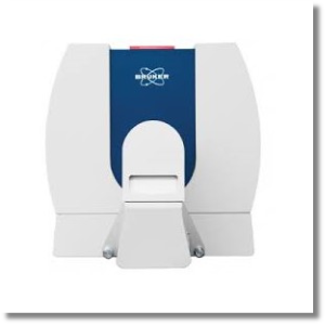
Since 2021, the Laboratory has also offered the possibility of performing optical imaging measurements in bioluminescence or fluorescence modes in the visible and near-infrared range. Compared to other techniques, optical imaging is characterized by extremely high sensitivity to molecular and metabolic processes and can identify events affecting individual cells. Some of its classic applications include tracking the biodistribution of cells labeled with fluorescent reporters and studying enzymatic reactions and gene expression involved in disease progression.
Equipment
Rates
The Centre’s regulations provide for the application of a fee for the use of the equipment:
INTERNAL USERS
Consultation is reserved for University staff: link
EXTERNAL USERS
| Equipment/Service | Autonomous use (hourly rate) | With technical support (hourly rate) |
|---|---|---|
| µCT ScyScan 1276 | not available | € 110,00 |
| MRI Bruker PharmaScan | not available | € 140,00 |
| IVIS Lumina III Perkin Elmer | not available | € 50,00 |
| Anesthesia | not available | € 5,00 |
| Special treatments of the sample, organization and treatment of data, drafting of reports | not available | € 100,00 |
EXTERNAL USERS WITH AGREEMENTS
| Equipment/Service | Autonomous use (hourly rate) | With technical support (hourly rate) |
|---|---|---|
| µCT ScyScan 1276 | € 37,20 | € 67,20 |
| MRI Bruker PharmaScan | € 57,60 | € 87,60 |
| IVIS Lumina III Perkin Elmer | € 4,80 | € 32,40 |
| Anesthesia | € 5,00 | € 5,00 |
| Special treatments of the sample, organization and treatment of data, drafting of reports | not available | € 30,00 |

