

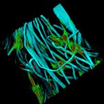
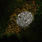
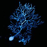
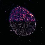
Optical Microscopy
"A picture is worth a thousand words", said Stefan Hell, Nobel Prize winner in Chemistry and father of nanoscopy, opening his lecture on 8 December 2014 in Stockholm. And this has been true for the life sciences since the invention of the microscope.
Description
The Optical Microscopy Laboratory provides support and assistance to users from experiment design and image acquisition to quantitative data analysis.
Our wide range of cutting-edge technologies — some of which can be used independently after a dedicated training — and our advanced techniques allow us to answer a variety of scientific questions.
The laboratory is equipped with wide-field microscopes, as well as optical sectioning and nanoscopy setups for acquiring images of fixed and living samples.
A sample preparation room (BSL2) and a workstation dedicated to image analysis are also available.

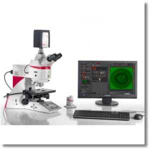
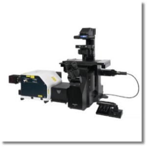
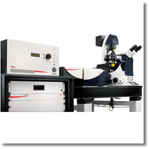
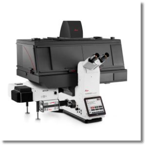
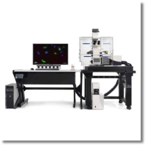
Equipment
Software
| Nome | Descrizione | |
|---|---|---|
| Zeiss arivis Pro Software | Zeiss arivis Pro is a comprehensive software solution for viewing, sharing, analyzing and presenting multi-channel and multi-dimensional (2D, 3D, and 4D) image data. The software supports and manages over 30 commercial file formats, efficiently processing even large files. Arivis Pro enables the analysis of even complex models using standard or AI model-based pipelines and enables workflow integration via Python scripting. | link |
| SVI Huygens Deconvolution Software | Huygens is a custom-built software for deconvolution and processing of microscopy images, capable of deconvolving a wide range of images (widefield, confocal, light-sheet, STED). Its user interface guides the user through the image deconvolution process, allowing comparison between raw and deconvolved results and multidimensional rendering of the data. | link |
| Open source Image Processing Software | CellProfiler, ImageJ/Fiji, Leica LasX Lite. |
Rates
The Centre’s regulations provide for the application of a fee for the use of the equipment:
INTERNAL USERS
Consultation is reserved for University staff: link
EXTERNAL USERS
| Equipment/Service | Autonomous use* (hourly rate) | With technical support (hourly rate) |
|---|---|---|
| STED/DSL/TIRF/TCS SP8 microscopes | not available | € 150,00 |
| SPINNING DISK microscope | not available | € 150,00 |
| DM6 microscope | €25,00 | € 50,00 |
| Time Lapse | €7,00 | not available |
| Data analysis | not available | € 100,00 |
| Special treatments of the sample, organization and treatment of data, drafting of reports | not available | € 100,00 |
* autonomous use permitted upon proven competence in using the equipment
EXTERNAL USERS WITH AGREEMENTS
| Equipment/Service | Autonomous use* (hourly rate) | With technical support (hourly rate) |
|---|---|---|
| STED/DSL/TIRF/TCS SP8 microscopes | not available | € 60,00 |
| SPINNING DISK microscope | not available | € 36,00 |
| DM6 microscope | not available | € 9,60 |
| Data analysis | not available | € 30,00 |
| Special treatments of the sample, organization and treatment of data, drafting of reports | not available | € 30,00 |
* autonomous use permitted upon proven competence in using the equipment
Publications
FAQ and Protocols
FAQ
What types of sample support can be used for experiments on our instruments?
Please contact the lab manager to verify the compatibility of your supports before proceeding with the experiment.
Why do slides need to be mounted and which mounting solution is right for my slides?
The choice of mounting medium largely depends on the type of sample being examined. Numerous mounting solutions exist, which are generally based on organic solvents, such as toluene or xylene, or aqueous solutions. These are available ready-to-use or can be prepared in the laboratory. Aqueous solvents are the most commonly used for sample preparation in optical microscopy. They are divided into two classes depending on whether they solidify or not. The advantage of polymerizing mounting media is that they can be stored for extended periods, even at -20°C, without losing their ability to preserve existing stains. However, they solidify within a few days, which leads to sample crushing. Therefore, they are not the best choice for imaging purposes such as 3D reconstruction of the sample structure or protein colocalisation analysis. In these cases, samples should be mounted using a liquid solution, such as a glycerol-based one. While the 3D structure remains unaffected, the stains are preserved for much shorter periods, only few days. The edge of the coverslip must also be fixed to the slide using VALAP or nail polish.
Is it possible to perform analyses on living samples or to perform live cell imaging experiments over time in the Optical Microscopy Laboratory?
Is it possible to perform Fluorescence Recovery After Photobleaching (FRAP) experiments in the Optical Microscopy Laboratory?
Where can I find information about the excitation and emission spectra of fluorophores?

