

Flow Cytometry
"Cytometry reflects the beauty of the world one cell at a time." - Dorothy E. Lewis
Description
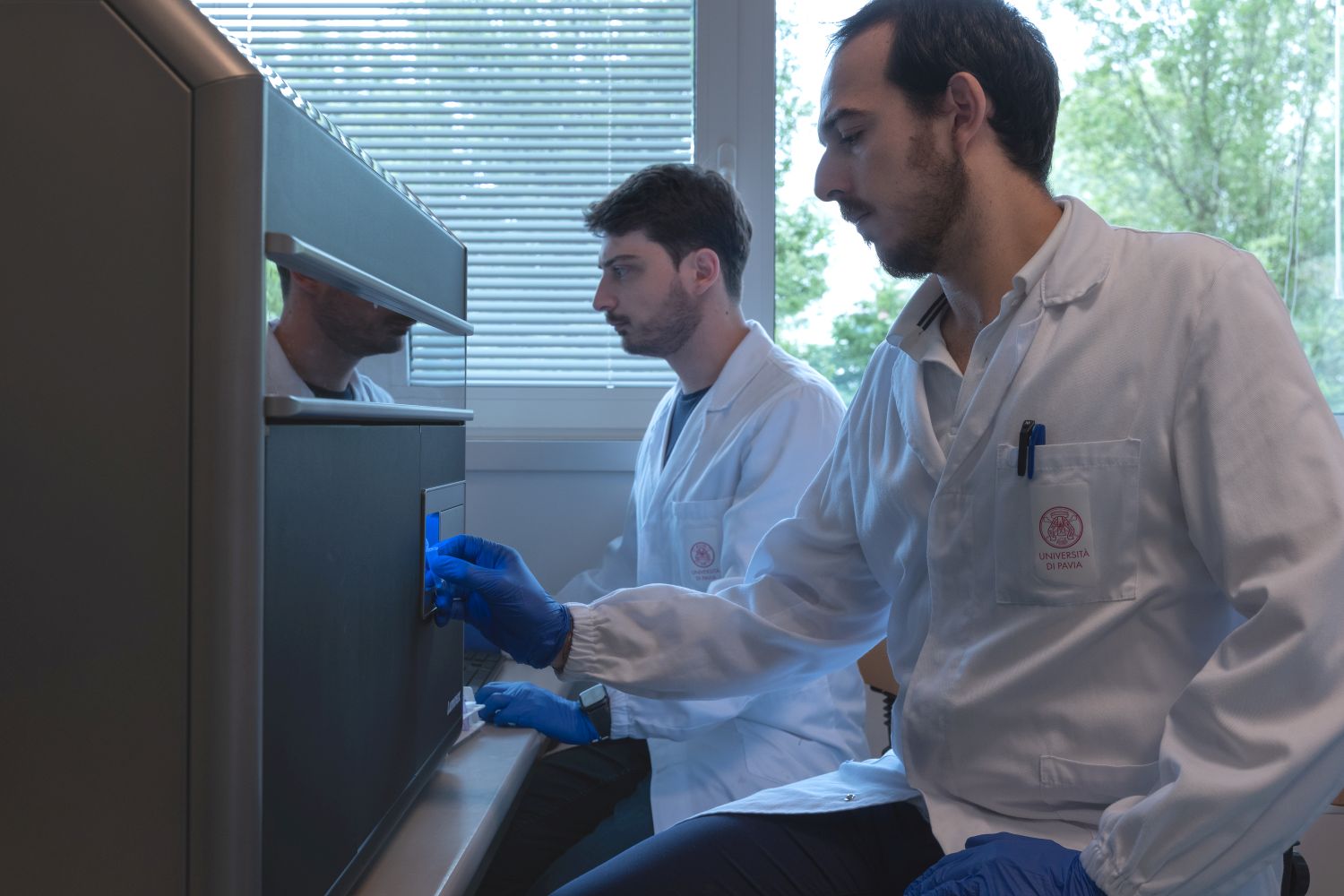
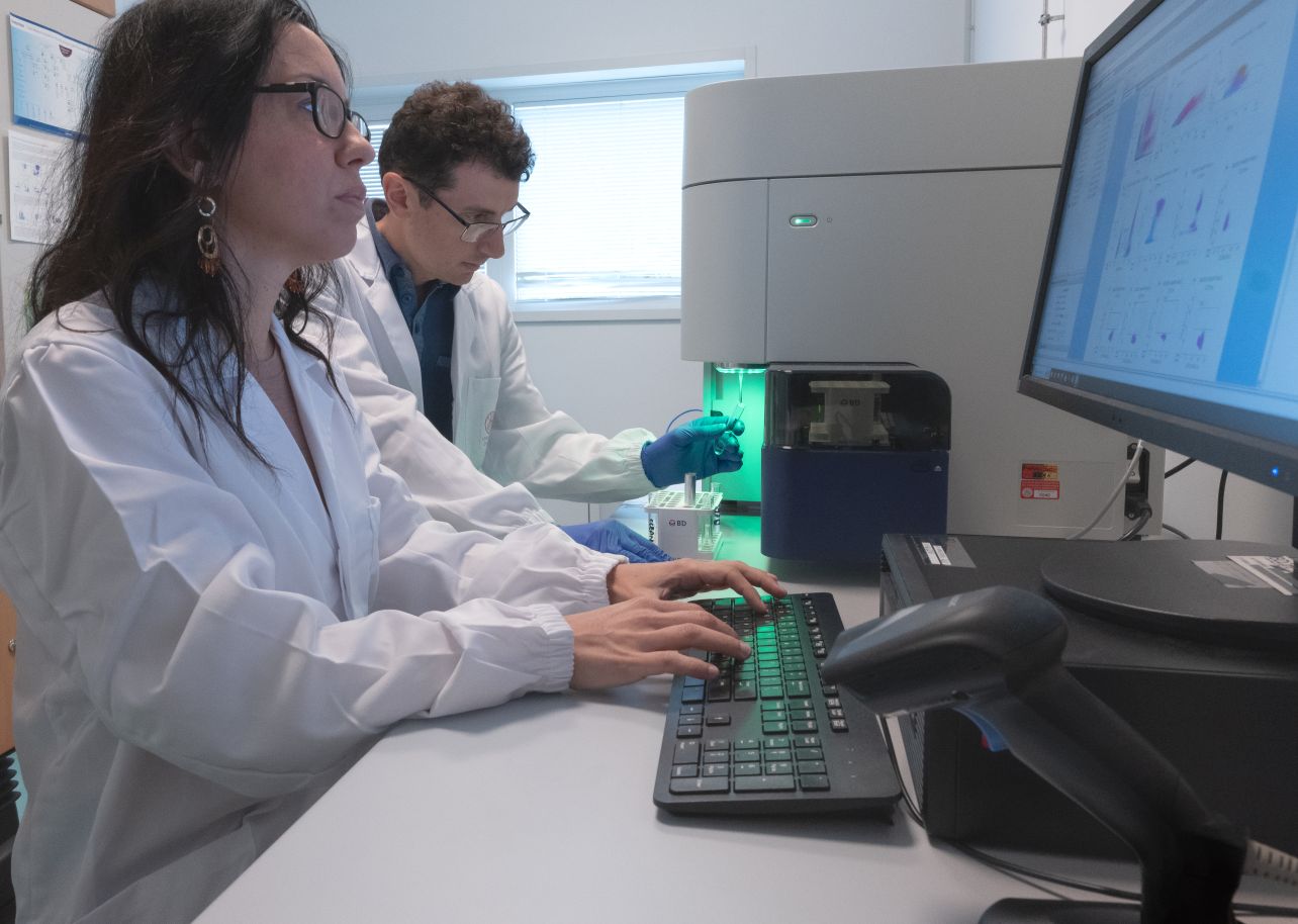
The Flow Cytometry Laboratory offers support and assistance to both internal and external users at different levels.
LEVEL 1 – Training and Education
Technical and Analytical Skills
- Hands-on training on instruments: flow cytometer (BD), imaging cytometer (Amnis), cell sorter (BD)
- Training on different available software for data analysis: FACSDiva, FacsSuite, IDEAS (Amnis), FlowJo
- Basic and advanced theoretical courses
LEVEL 2 – Scientific Assistance
Expert support for complex experiments
- Assistance with multiparametric panels (phenotyping)
- Assistance with multiparametric sorting panels (sorting with different masks: purity, yield, single cell)
LEVEL 3 – Accessibility and Collaboration
Open to internal and external users
- Personalized technical and scientific support
- Collaboration on complex projects
The lab is equipped with three flow cytometry instruments, a BSL-2 sample preparation room, and a dedicated flow cytometry analysis workstation.

Equipment

Software
| Nome | Descrizione | |
|---|---|---|
| FACSSuite | The software that controls the BD FACSLyric™ flow cytometer consists of two applications: BD FACSuite™ Clinical Application and BD FACSuite™ Application. Both provide a user-friendly interface for data acquisition and analysis with the BD FACSLyric™ system. The BD FACSuite™ Clinical Application supports IVD diagnostic testing, while the BD FACSuite™ Application is designed for custom, user-defined tests, offering a simple, reproducible, and transferable configuration that facilitates standardization. | link |
| FACSDiva | It is a comprehensive system that provides tools for cytometer setup, application configuration, data acquisition, and result analysis. It is designed to streamline flow cytometry workflows in modern, high-throughput laboratories. BD FACSDiva™ offers features that help users integrate flow cytometry systems across various applications, including index sorting and single-cell applications. It provides practical and intuitive tools for building experimental layouts, simplifying experiment setup. Users can visually navigate the entire experiment and quickly add labels, keywords, and acquisition attributes to a single tube, multiple tubes, specific samples, or the entire experiment. The software allows users to save templates of experiments, analyses, and panels for future use—without compromising data integrity. It also enables saving application-specific settings to correctly identify cell populations of interest. Once a daily performance check is completed to update these settings, they can be applied to ensure consistent execution of applications day after day. | link |
| FlowJo | The most widely used software for flow cytometry data analysis is available in the facility on a dedicated workstation with a portal licence. Key features: Advanced data analysis: enables the analysis of large data sets, including dozens of parameters per cell. Dynamic gating: allows users to define and refine cell populations in a visual, interactive, and precise manner. Statistics and plots: calculates statistics for each population (mean, standard deviation, percentages, etc.) and generates various plots such as histograms, dot plots, and density plots. Plugins: includes modules for advanced analyses such as kinetics, cell cycle, apoptosis, automated clustering, t-SNE, UMAP, SPADE, and index sorting. Compatibility: supports .fcs files, the standard format for flow cytometry data, and is compatible with data from various instrument manufacturers (BD, Beckman Coulter, Sony, Cytek, etc.). Batch analysis: allows automated analysis of dozens or hundreds of samples using the same gating strategy. | link |
| Inspire | It is a data acquisition software developed by Amnis for use with the ImageStreamX Mark II flow cytometer. This instrument combines the capabilities of traditional flow cytometry with high-resolution imaging, enabling simultaneous acquisition of brightfield and fluorescence images of cells in flow. It supports high-speed acquisition, capturing both brightfield and darkfield images, and manages image capture via a TDI CCD camera, ensuring high spatial resolution (up to 0.3 × 0.3 µm per pixel with a 60× objective). Acquired data can be analysed using IDEAS software, which provides quantitative tools for cell image analysis and population-level statistics. This system is used to study various cellular processes such as: translocation, co-localization, cell cycle, apoptosis, autophagy, vesicle internalization. | |
| IDEAS | It is the data analysis software developed by Amnis, specifically designed for processing data generated by ImageStream imaging flow cytometers. It enables the analysis of images acquired through imaging flow cytometry by integrating fluorescence intensity with morphological information, allowing for the quantification of complex cellular events such as: co-localization, nuclear translocation, nuclear morphology, apoptosis, phagocytosis, internalization. The software also allows users to create custom masks to identify subcellular regions (e.g. nucleus, cytoplasm, membrane) for the calculation of statistical measurements on those specific areas. |
Rates
The Centre’s regulations provide for the application of a fee for the use of the equipment:
INTERNAL USERS
Consultation is reserved for University staff: link
EXTERNAL USERS
| Equipment/Service | Autonomous use* (hourly rate) | With technical support (hourly rate) |
|---|---|---|
| BD FACSLyric Flow Cytometer | € 30,00 | € 60,00 |
| AMNIS IMAGE STREAM MARK II Flow Cytometer | € 45,00 | € 90,00 |
| BD FACSAria III Cell Sorter | € 40,00 | € 80,00 |
| Special treatments of the sample, organization and treatment of data, drafting of reports | not available | € 100,00 |
* autonomous use permitted upon proven competence in using the equipment
EXTERNAL USERS WITH AGREEMENTS
| Equipment/Service | Autonomous use* (hourly rate) | With technical support (hourly rate) |
|---|---|---|
| BD FACSLyric Flow Cytometer | € 13,20 | € 40,70 |
| AMNIS IMAGE STREAM MARK II Flow Cytometer | € 13,20 | € 40,70 |
| BD FACSAria III Cell Sorter | € 39,60 | € 67,10 |
| Data analysis using Flowjo software | € 4,00 | not available |
| Special treatments of the sample, organization and treatment of data, drafting of reports | not available | € 30,00 |
* autonomous use permitted upon proven competence in using the equipment
Publications
FAQ and Protocols
FAQ
When should I use FACS (Fluorescence-Activated Cell Sorting) instead of other separation methods, such as density gradient centrifugation or immunomagnetic separation?
- When high purity (greater than 95%) of the target cell population is required.
- For separations based on monochromatic or polychromatic staining using fluorescent dyes or fluorophore-conjugated antibodies.
- For separations based on intracellular staining, such as DNA or internal antigens.
- For the separation of populations with low surface receptor density.
- For the enrichment of cell populations based on surface receptor density.
How large can the cells be?
How many cells do I need for the sorting?
To answer this question, the following information is required:
- What is the percentage of the target cell population?
- How many cells do you need after sorting?
- What is more important: high purity or overall yield?
- How fragile or large are the cells?
The duration of the sorting procedure depends on all these parameters.
Here is an example:
How many cells should I prepare to obtain 1×10⁶ cells of a population that represents 10% of the sample?
1×10⁶ = 0.1 [10% target population] × 10×10⁶ starting cells.
However, the actual yield is typically around 80–90% of this theoretical value.
Therefore, the starting number of cells should be:
10×10⁶ × 100 / [80–90] = 11–12.5×10⁶ cells.
The yield can be significantly lower if the cells are of poor quality (low viability, aggregation, etc.) or if high purity is required.
It’s also important to remember that a portion of the sample will be used to set up the sorter.
If it’s your first time sorting a particular sample, you may need more cells than in subsequent experiments.
What does the yield of a sorting depend on?
The actual recovery percentages depend on several factors:
- Cell quality: this is the most critical factor affecting yield. Cells must be free of aggregates and in a single-cell suspension.
- Cell death before and after sorting, or loss due to adhesion to the tube walls.
- Sorting speed: the higher the speed, the lower the yield.
- Accuracy of the sorter setup.
- Whether the sorting is for enrichment or for purity: enrichment sortings typically result in higher yields.
- Losses during sample preparation.
When a user claims to have brought a sample with 1×10⁷ cells, this number should reflect the cell count after the entire preparation process, including filtration to remove aggregates.
Sorters accurately count the cells that pass through the instrument.
Therefore, if the user assumes to have brought 1×10⁷ cells but the sorter detects only 0.6×10⁷, then the actual number of cells is not 1×10⁷.
It should also be noted that a small aliquot of the sorted cells can be used to assess sorting efficiency. However, in cases of very low yield, this verification may be skipped.
That said, post-sort analysis is essential if you plan to publish the results of the experiment.
Tip: use at least 50% more cells than the theoretical number required for sorting.
What percentage of the theoretical number of cells can realistically be recovered? What does it depend on?
Losses are caused by the factors already discussed in FAQ 4.
Are there ways to improve sorting?
- Use polypropylene tubes for cell preparation and sorting.
- Use polypropylene collection tubes and fill them (about 1/2 mL) with medium containing 30% serum (for eukaryotic cells).
- Count the cells immediately before sorting (after all washes).
- Use the “Yield” sorting mode to improve recovery (Note: purity will decrease!).
- Process collection tubes immediately after filling or at the end of sorting.
How many cells per second can be sorted with a BD FACSAria III sorter?
For certain cell preparations, lower speeds—such as 5,000 events per second or less—may be recommended to achieve higher purity.
How long does the sorting procedure take?
Approximately 1 hour is required for each working day for the operator to prepare the instrument, stabilize the fluidics, and complete quality control (QC) checks.
This is followed by 15–45 minutes to set up the sorting strategy, and about 15 minutes for post-sort analysis.
The actual sorting duration mainly depends on how many cells need to be analysed and sorted.
Theoretically, the BD FACSAria III can process up to 20–30 million events per hour.
Other factors that influence the overall duration include:
- The quality of the cells.
- The percentage of the target population.
- The number of cells needed after sorting.
- Whether yield or purity is selected.
- The concentration of the sample.
The more diluted the cells are, the more time is required.
For dilute samples, volume also can affect the time of the experiment. Typically, 1 mL of sample (up to 20–30 million cells) can take about 2 hours to process.
Will my cells be damaged during the sorting?
In general, cells may experience mechanical stress during the process, but this can be minimized by keeping them at the appropriate temperature, pH, and in the most suitable medium.
The BD FACSAria III is equipped with a 100 μm nozzle, which can be operated at lower pressure compared to smaller nozzles, automatically reducing mechanical stress.
Additionally, the user can set the temperature of the collection chamber, helping to maintain optimal conditions for the cells.
How can I preserve cell viability during sorting, and are there ways to improve it?
For eukaryotic cells, it is advisable to add serum proteins, but without exceeding a concentration of 2–3%, as a high serum content in the sample may compromise sorting accuracy.
For eukaryotic cell lines and adherent cells, adding 0.5 mM EDTA is recommended. For particularly sticky cells, the concentration can be increased up to 5 mM.
For sterile sorting, the addition of antibiotics is recommended. It can also be helpful to include 25 mM HEPES at pH 7.0.
Collection tubes should contain a high serum concentration (30%), as this will be diluted by the sheath fluid carried by the sorted droplets.
Processing the cells immediately after sorting helps maintain their viability.
What is the maximum purity that can be achieved for a sorted population, and what does it depend on?
A purity of 95%–100% can be expected for populations that are well-separated from unwanted cells.
Purity depends on the stability of the sorter and how the sorting gates are set.
For poorly defined regions or “enrichment”-type sorting, purity may drop below 90%.
How is the purity of the sorted population determined?
In the latter case, make sure the sensitivity of the instruments is comparable.
What should the cell concentration be for the sorting?
- If there are too few cells, sorting will take longer and cell viability may be affected.
- If the cells are too concentrated, it may reduce purity and cause instrument clogging.
👉 An optimal cell preparation improves viability and leads to higher yield.
Guidelines:
- If you have fewer than 10×10⁶ cells, prepare them in a volume of 0.5 mL.
- The recommended concentration for the BD FACSAria III is 20–30 million/mL, depending on cell type.
- If the sample is too concentrated, we can dilute it at the facility.
- If unsure, it’s better to bring the sample more concentrated (it can always be diluted).
- Count the cells right before sorting (after all washes).
What buffer should cells be prepared in?
- In general, any physiological buffer can be used, but it is essential to avoid cell clumps (aggregates).
- For prokaryotic cells: PBS (phosphate buffered saline).
- For eukaryotic cells: PBS or HBSS without Ca²⁺/Mg²⁺ and with ≤2% FBS or BSA. Standard media for eukaryotic cultures contain Ca²⁺, Mg²⁺, and serum proteins, which promote aggregation. Many cells benefit from the presence of proteins in the buffer (e.g., 2% FBS or BSA), but concentrations >5% can lead to aggregation and clogs.
- For cell lines and adherent cells, the addition of 0.5 mM EDTA is recommended, which can be increased to 5 mM for highly adhesive cells.
- In suspensions with a high percentage of dead cells, free DNA can coat the cells and cause severe clumping: adding DNase II (20–100 μg/mL; 10 U/mL) may help.
- For cells sensitive to pH, adding HEPES (10–25 mM) is advisable to buffer pH fluctuations caused by high pressure.
- The addition of antibiotics is recommended for sterile sorting conditions.
What is the recommended resuspension volume for the sorting?
- The minimum recommended volume is 0.4–0.5 mL.
- The maximum volume that can be processed in a single run is 3–4 mL.
What should be considered during cell preparation?
For example, large (eukaryotic) cells should be filtered—ideally right before sorting—using cell strainers of 20–50 µm.
What tubes are required for cell sorting?
Additional collection tubes can be prepared if needed.
How should I transport the cells?
What types of tubes are used for cell collection?
Please bring labeled collection tubes containing about 1/2 mL of cell culture medium.
Sterile 5 mL polypropylene FACS tubes are also required for post-sort acquisition controls.
Additionally, the BD FACSAria III is equipped with a holder for single-cell collection into 96- (and 384-) well plates. Plates should be brought pre-filled with medium.
Which fluorochromes can be analysed?
It is necessary to verify in advance whether the fluorescent proteins or fluorochromes used can be detected by the cytometer.
Not all fluorochromes visible under the microscope can also be measured by the cytometer.
To check if the optical configuration is suitable, online tools called Spectrum Viewers are available from various companies, for example:
- https://www.bdbiosciences.com/us/s/spectrumviewer
- https://www.thermofisher.com/us/en/home/life-science/cell-analysis/labeling-chemistry/fluorescence-spectra-viewer.html
The configuration of the BD FACSAria III is provided in the Equipment section.
Is it possible to sort single cells into plates?
What is index sorting?
How many parameters can be used simultaneously for the sorting strategy on the BD FACSAria III?
The UV and violet lasers cannot be used simultaneously in the same experiment.
Which controls should I bring with me?
It is recommended to bring the following controls for flow cytometry experiments:
- Unstained cells as a negative control.
- Unstimulated/untreated cells as a biological control.
- Single-stained cells or beads for compensation.
- Isotype control or FMO (fluorescence minus one) control.
- If using a viability dye to exclude dead cells, bring a separate viability control for proper gating, since dead cells cannot always be excluded based on light scatter alone. It is recommended to kill part of the cells, mix them with live cells, and then stain with your viability dye (e.g., PI, 7-AAD, DAPI, TO-PRO-3).
- An untransfected control in experiments with fluorescent proteins.
Why is it important to remove dead cells during sorting of eukaryotic cells?
Dead cells represent a significant issue in cell sorting. Generally, if a suspension contains more than 10% dead cells, they must be removed. Below this threshold, sorting performance may still be negatively affected.
The main reasons to eliminate dead cells are:
- Dead cells release free DNA, which can cause cell clumping.
- Dead cells can bind antibodies nonspecifically, leading to false positives.
The first problem can be mitigated by adding DNase I.
The second issue worsens especially in antibody-mediated separations, as dead non-target cells may still bind antibodies, compromising purity.
The most effective solution is to completely remove dead or dying cells via a pre-sorting purification step.
Is sorting performed under sterile conditions?
The BD FACSAria III sorter is not placed inside a Class II laminar flow hood, so no sorting can be considered absolutely “aseptic”.
However, sorting is carried out aseptically:
- The sheath fluid passes through a 0.22 µm filter.
- The sorter is regularly decontaminated (the day before and after each session) with FACSClean™ and sterilized with 70% ethanol.
It is the user’s responsibility to prepare samples under sterile conditions (it is recommended to keep a pre-sort control aliquot to verify sterility). All materials (buffers, media, antibodies, tubes, tips) must be sterile.
For sorted eukaryotic cells, the use of antibiotics in the medium is recommended.
Our facility is equipped with a Class II biological laminar flow hood for sample filtering.
Which other preparations are required?
Please bring a recent printout of the data at your first appointment with the staff.
Punctuality is recommended!
How is data storage managed in the FACS service?
Acknowledgments and co-authorship
We kindly ask you to send us a PDF of accepted articles containing data generated by our service and to inform us if your funded projects include data obtained through the FACS service.
For more information, please refer to the policy.
Protocols
- Sorting Guidelines: Planning an Experiment (link)
