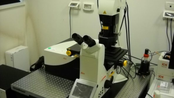
Point scanning confocal microscope, equipped with a 405nm Diode laser, the White Light Laser, tunable from 470nm to 670nm, and a 488nm AR laser.
The White Laser allows for extremely precise selection of the excitation wavelength for each fluorophore: optimizing excitation efficiency reduces fluorophore photobleaching and sample photodamage.
The system is equipped with four detectors, including two hybrid detectors, a top-stage incubator for live cell imaging and the AFC (Adaptive Focus Control) hardware focus control tool.
The Navigator tool for acquiring specific regions of the sample (mosaic and multipoint acquisition) and a 3D rendering tool are also available as well as the Resonant Scanner module, essential for tracking fast cellular processes in live cell imaging, but also for optimizing acquisition time of large, fixed samples.
The microscope is equipped with the DLS (Digital Light Sheet) module for fast and sensitive acquisition of thick, naturally transparent or clarified samples, both fixed and in living conditions (organoids, spheroids, Drosophila and zebrafish embryos, tissue sections, and small organ parts). The samples must be housed in glass capillaries with a maximum diameter of 2.5 mm.
Available objectives:
- HC Pl Apo 20X/0.75 imm CS2;
- HC Pl FLUOTAR (340) 40X/1.30 oil;
- HC Pl Apo 63X/1.40 oil CS2;
- Fluotar VISIR 25X/0.95 water.
DLS available objectives:
- Illumination objectives: 1.6X/0.05, 2.5X/0.07;
- Detection objectives: 5X/0.15 imm incl BABB, 10X/0.3 W, 25X/0.95 W;
- Mirrors: 2.5mm imm, 5mm imm, 7.8mm W, 7.8mm Gly, 7.8mm BABB.
Available techniques:
- Multi-channel analysis;
- Colocalization analysis;
- 3D reconstruction;
- Tile-scan imaging (Navigator);
- Spectral unmixing;
- Lambda-scan (reconstruction of excitation and emission spectra);
- Time-lapse;
- FRAP (Fluorescence Recovery After Photobleaching) and photoperturbation techniques;
- FRET (Fluorescence Resonance Energy Transfer);
- Laser microirradiation to produce localized DNA damage;
- Lightning tool (deconvolution);
- DLS.

