
Marta Filibian
tel. +39038298464 marta.filibian@unipv.it
The multimodal platform perform advanced Imaging studies and sequential analyses of the same sample by means of both Magnetic Resonance Imaging (MRI) and µCT (Computed Microtomography). Notably, this approach offers the unique advantage to directly compare the images obtained by the two techniques, using the same software.
MRI allows to acquire a wide range of morphologic, metabolic and functional information from the nervous system and several other organs and tissues. In particular, the application of this technique is crucial for non invasive studies of tumors, edema, lesions, of the hearth (Cardio MRI), and for studies regarding the connectivity and the functions of the different areas activated within the nervous system (e.g. by means of Diffusion Tensor Imaging and Functional Imaging, fMRI). Moreover, using the MRI tomograph, it is possible to conduct Magnetic Resonance Spectroscopy (MRS) analyses, which retrieves the simultaneous and accurate quantification of several metabolites contained in tissues, such as N-acetylaspartate (NAA), Choline (Cho), Creatine (Cr), Myo-inosytol (mIns), Glutamate (Glu), Glutamine (Gln), Lactate (Lac), whose concetration can be severely altered in presence of pathologies , e.g. neurodegenerative diseases and tumors.
X-ray Computed Microtomography (microCT) provides the 3-dimensional internal structure of samples with a minimum pixel size of 2.8 μm. The 3D structure is the result of a complex reconstruction, obtained from a set of 2-dimensional shadow images of samples, produced by the X rays passing through them. MicroCT is optimal for the study of bones, to obtain highly detailed 3D reconstructions without compromising the subsequent genetic and histological analyses. The application of this technique, with the simultaneous usage of contrast agents, also allows non invasive investigation of lungs, of the cardiovascular system, of cartilages and other soft tissues. The microCT scanner can be also used for the structural studies of biomaterials and biopolimers.
Equipment
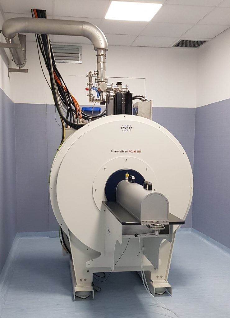
Bruker PharmaScan MRI scanner equipped with an horizontal 7 Tesla Magnet, with a bore of 16 cm diameter. The Pharmascan system is based on Avance NEO electronics and works with Bruker BGA 9S HP gradients yielding a maximum strength of 760 mT / m. The scanner has 4 channels for transmission and reception of signals and can detect both protons and X nuclei such as 13C, 19F, 23Na. The samples can be located inside the magnet bore in a very precise and reproducible way with the ManPac system. The high magnetic field of the PharmaScan increases the sensitivity of MRI studies and allows the investigation of millimetric structures with a suitable resolution. Moreover, it also improves the sensitivity in other advanced applications depending on the Chemical Shift, such as MRS, and from local Susceptibility, such as the fMRI with BOLD (Blood Oxygenation Level Dependent) contrast
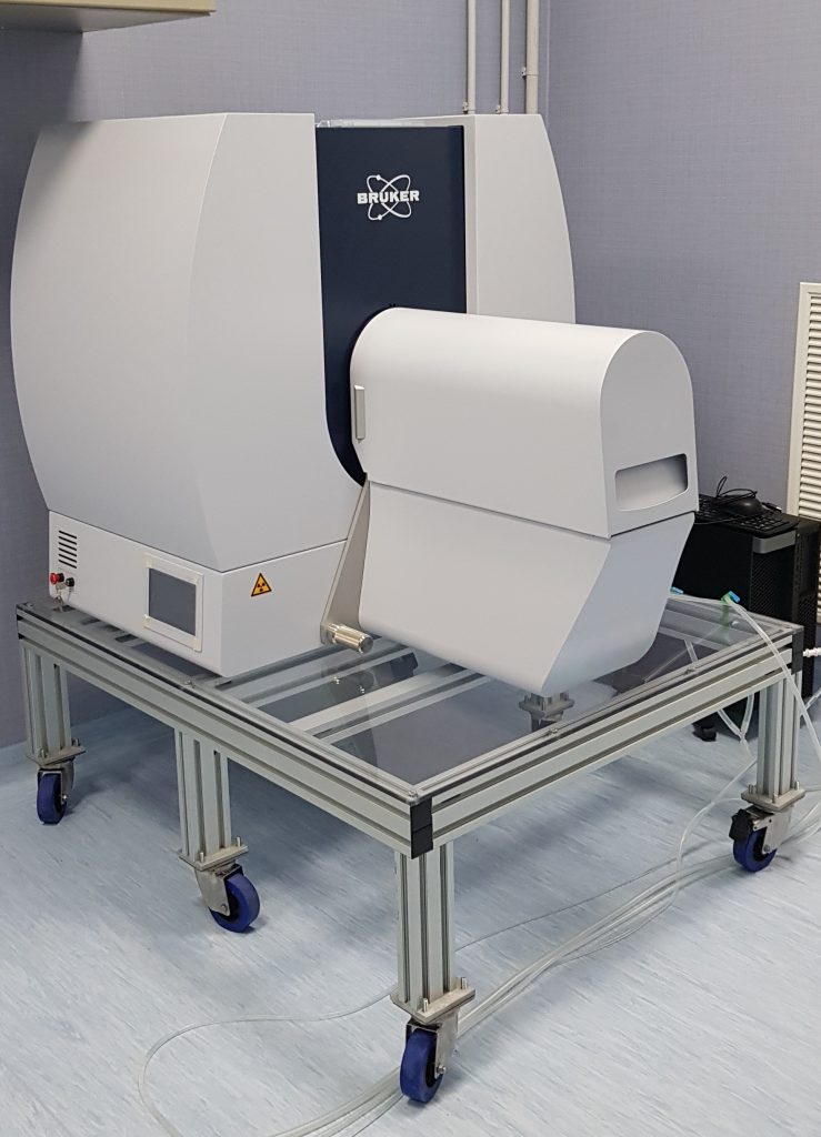
Skyscan 1276 system equipped with a 20-100 kV micro-focus X- ray source and a 11 Mp detector. It can apply variable magnification until reaching a high resolution with minimum pixel size below 3 Microns, enough to resolve structural details of the order of 5-6 Microns. The SkyScan is particularly indicated for longitudinal studies, because the radiation dose can be properly limited. The image analysis is performed with a dedicated software, InstaRecon, capable of providing fast reconstruction of 3D realistic images