
Andrea Alfieri
tel. 0382.985525 andrea.alfieri@unipv.it
Cryoelectron microscopy (cryo-EM) allows scientists to determine the structures of biological macromolecules (proteins, nucleic acids and their complexes) at near-atomic resolution. The sample is purified and vitrified in electron microscopy grids using liquid ethane and liquid nitrogen. No colouring or fixation is needed. The technique maintains the sample in a paraphysiological aqueous environment, where multiple native conformations of the macromolecules can be visualized even simultaneously. The acquisition of hundreds of thousands of 2D projections of a particle allows to reconstruct its 3D structure by computational methods (“single particle analysis”
Equipment
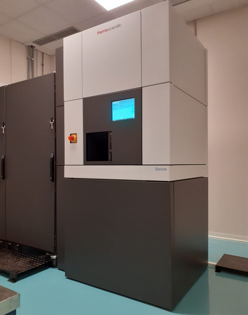
- Cryo transmission electron microscope Glacios (Thermo Fisher Scientific) equipped with 200 kV high-tension generator, X-FEG electron source, liquid nitrogen cooling, sample autoloader, CMOS detector (Ceta 16M Camera) and direct electron detector (Falcon 3EC)
- Satellite instrumentation: cooling unit (in a separate room); network PC for data storage (in a separate room)
- Vitrobot (Thermo Fisher Scientific) device for PC-controlled vitrification of aqueous samples
- Instrumentation for preparing and manipulating electron microscopy grids at cryogenic temperature